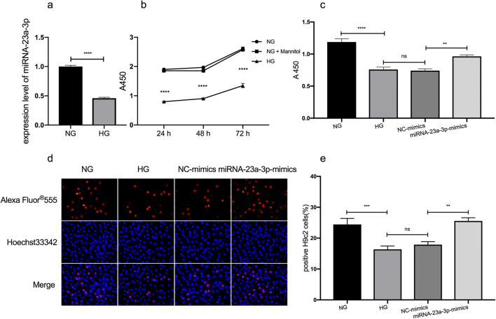
Figure 1.
Effects of miRNA-23a-3p overexpression on cell proliferation in HG-treated H9c2 cells. (a) RT-qPCR to detect the expression level of miRNA-23a-3p in HG-treated H9c2 cells (****p < 0.0001 vs. NG group). (b) H9c2 cells were exposed to glucose at concentrations of 5 mM (NG), 35 mM (HG), and 5 mM glucose + 30 mM mannitol (NG + Mannitol) for 24, 48, and 72 h (****p < 0.0001 vs. NG group). Cell viability was assessed by a CCK-8 assay. (c) H9c2 cell viability was significantly increased following treatment with the miRNA-23a-3p mimics (**p < 0.01 vs. NC-mimics). (d) Representative images illustrating the EdU and Hoechst staining of H9c2 cells exposed to 5 mM and 35 mM glucose and transfected with NC mimics or miRNA-23a-3p mimics. (e) Analysis of EdU-positive cardiomyocytes by an EdU incorporation assay. The percentages of proliferative H9c2 cells were calculated (n > 500). Compared with the NC-mimics group, the miRNA-23a-3p-mimics group had increased EdU incorporation (**p < 0.01). CCK-8 Cell Counting Kit-8, EdU 5-ethynyl-2′-deoxyuridine, NG normal glucose, HG high glucose, NC negative control.

