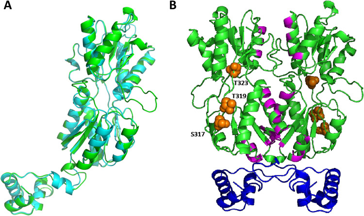
FIG 1.
Structural homology model of S. pneumoniae GalR. (A) Cartoon representation of the protomeric homology model of S. pneumoniae GalR (green) based on the 2.9-Å structure of the Escherichia coli PurR W147F mutant (cyan; RMSD, 2.868 Å). (B) Cartoon representation of the dimeric homology model of GalR. The DNA binding helix-turn-helix domain is shown in blue, and the putative sugar binding regions are highlighted in magenta. The serine (S317) and threonine (T319 and T323) residues hypothesized to be phosphorylated are depicted as orange spheres.

