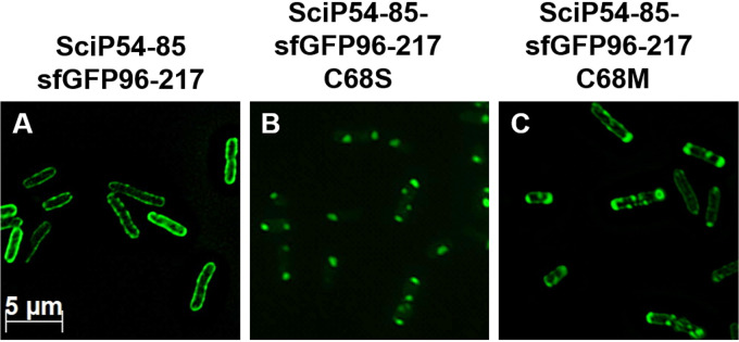
FIG 3.
Fluorescence microscopy of various SciP-sfGFP fusion proteins. E. coli MC4100 cells were transformed with the respective plasmid, and the expression was induced with 1 mM IPTG for 1 h at 30°C. (A) The fluorescent signal of SciP54-85 was clearly found at the membrane, indicating a targeting function of this region. (B) Replacement of the cysteine residue at position 68 with a serine residue led to distinct spots, probably due to the aggregated protein. (C) Replacement of the cysteine with a methionine residue resulted in a mixed phenotype with distinct spots, mainly at the cell poles and signals found at the membrane.

