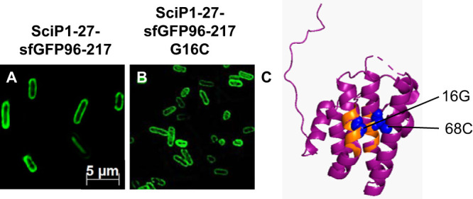
FIG 6.

(A and B) Subcellular localization studies of SciP1-27-sfGFP96-217 and the mutant fusion protein. The fluorescent signals of SciP1-27-sfGFP96-217 (A) and SciP1-27-sfGFP96-217 G16C (B) were clearly found at the membrane, showing that the mutation G16C has no influence on SRP-dependent targeting. (C) In the structure of the cytoplasmic domain of SciP, the two hydrophobic regions (amino acids 12 to 20 and 62 to 71) are colored orange, and the glycine residue at position 16, as well as the cysteine residue at position 68, are highlighted as blue spheres, respectively (image created with PyMOL 2.3.0; PDB ID 3U66).
