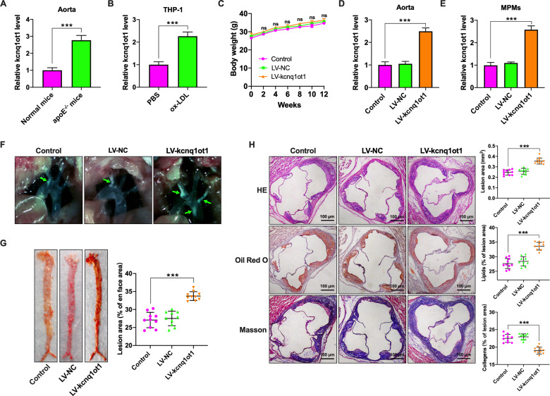
Fig. 1. Kcnq1ot1 aggravates atherosclerosis in apoE−/− mice.
A C57BL/6 mice and apoE−/− mice were fed a normal chow diet and a Western diet for 12 weeks, respectively (n = 10 per group). Kcnq1ot1 expression in the aorta was detected by qRT-PCR. B THP-1 macrophages were treated with PBS or 50 µg/mL ox-LDL for 48 h, followed by qRT-PCR analysis of kcnq1ot1 expression (n = 3). C–H Western diet-fed apoE−/− mice were injected via the tail vein with PBS, LV-NC, or LV-kcnq1ot1 (n = 20 in each group). C Comparison of weight. D, E The qRT-PCR analysis of kcnq1ot1 expression in the aorta and MPMs. F The plaques (green arrows) in the aortic arch of apoE−/− mice under a stereoscopic microscope. G The entire aorta was stained with Oil Red O and the atherosclerotic lesion area was quantified by analyzing Oil Red O-positive region on en face preparations (n = 5 per group). H Sections of the aortic root were stained with HE, Oil Red O, or Masson. Lesion area and percentage was quantified using Image-Pro Plus 7.0 software (n = 10 per group). Scale bar = 100 μm. Data are represented as mean ± SD. ***P < 0.001; ns not significant vs. control group.

