Figure 1.
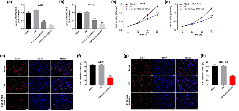
Knockdown of FGF12-AS2 significantly inhibited the proliferation of NSCLC cells. A549 or NCI-H23 cells were transfected with FGF12-AS2 shRNA1 or FGF12-AS2 shRNA2 for 24 h. Then, the expression of FGF12-AS2 in (a) A549 and (b) NCI-H23 cells was detected by q-PCR. (c and d) A549 or NCI-H23 cells were transfected with nothing, NC or FGF12-AS2 shRNA2 for 0, 24, 48, or 72 h. Then, the cell viability was detected by the CCK-8 assay. (e) The proliferation of A549 cells was tested by Ki-67 staining. Red immunofluorescence indicated Ki-67. Blue immunofluorescence indicated DAPI. (f) The positive rate of Ki-67 staining was calculated. (g) The proliferation of NCI-H23 cells was measured by Ki-67 staining. Red immunofluorescence indicated Ki-67. Blue immunofluorescence indicated DAPI. (h) The positive rate of Ki-67 staining was calculated. ** P < 0.01 compared to control.

