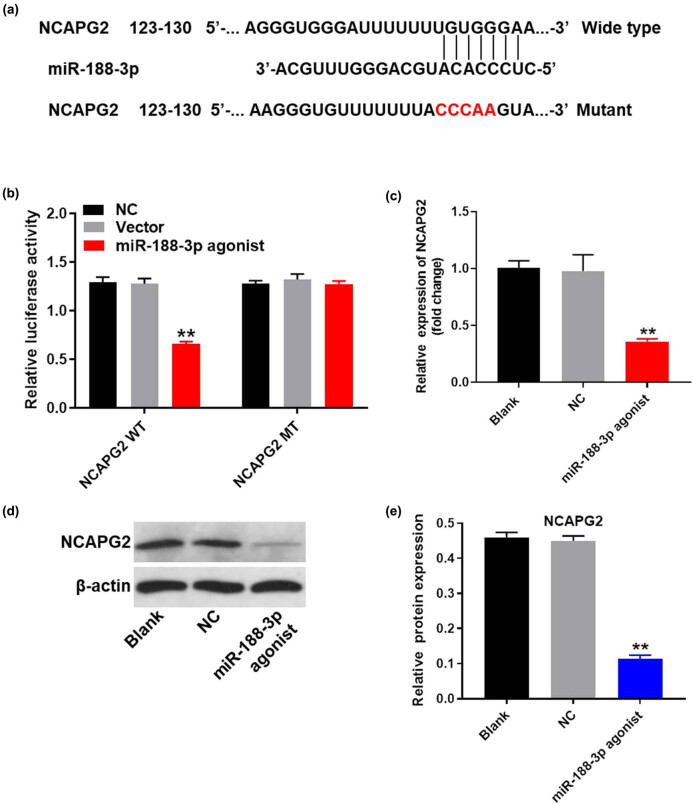
Figure 5.
MiR-188-3p directly targeted NCAPG2. (a) Gene structure of NCAPG2 at the position of 123–130 indicated the predicted target site of miR-188-3p in its 3′UTR, with a sequence of CCCAA. (b) The luciferase activity was measured in A549 cells following co-transfecting with WT/MT NCAPG2 3′-UTR plasmid and miR-188-3p with the dual luciferase reporter assay. (c) A549 cells were transfected with miR-188-3p antagonist for 24 h. The expression of NCAPG2 in A549 cells was detected by q-PCR. (d) The protein expression of NCAPG2 in A549 cells was measured by western blot. (e) The relative expression of NCAPG2 was quantified normalizing to β-actin. ** P < 0.01 compared to control.

