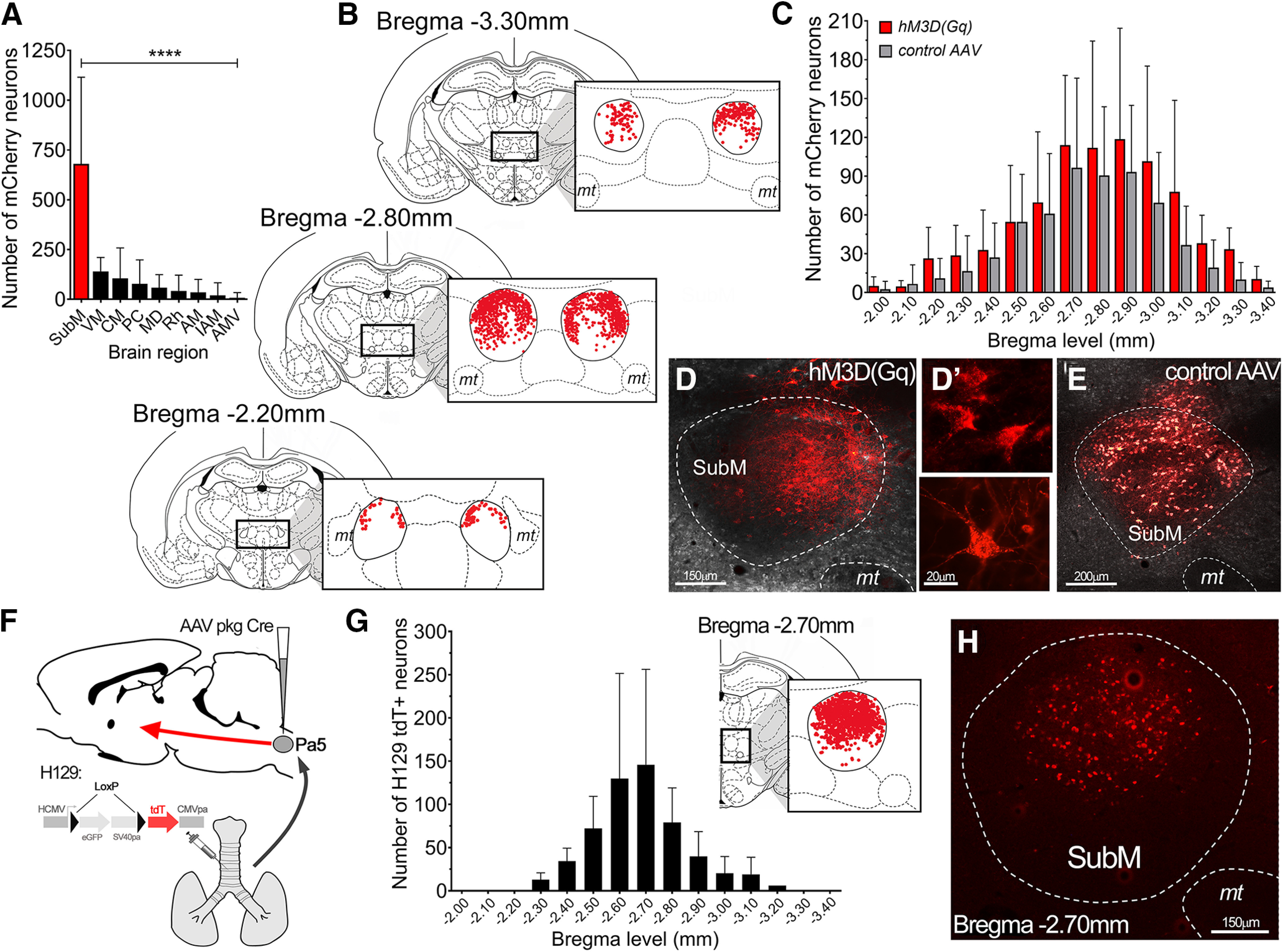
Figure 6.

Neuroanatomical organization of neurons in the submedius thalamic nucleus involved in modulating vagal reflexes. A, Total number of transduced neurons in the brain of hM3D(Gq) mCherry rats (N = 10). Data are presented as the mean ± SD for each brain region. B, Schematic representation of the distribution of VLO-projecting hM3D(Gq)-transduced neurons within the SubM. Each coronal slice summarizes the location of individual hM3D(Gq) mCherry neurons (red dots, from all hM3D(Gq) rats, N = 10) at three different bregma levels spanning the entire rostrocaudal extent of the SubM. C, Quantification of the rostrocaudal spread (at 100 µm intervals) of neurons transduced by hM3D(Gq) and control AAV (N = 9 rats). Data are represented as the mean ± SD. D–E, Representative images of hM3D(Gq) mCherry-transduced neurons (D) with examples of high-power individual neurons (D′) and control AAV mCherry-transduced neurons (E) in the SubM. F, Schematic representation of the anterograde transsynaptic HSV1-H129floxed viral tracer (permanently switches to tdTomato (tdT) fluorescence in the presence of Cre-recombinase; McGovern et al., 2015a) injected into the airways of rats that had previously been microinjected with the AAV pkg Cre construct into the medullary Pa5. G, Rostrocaudal spread of SubM H129 tdT-infected neurons in receipt of Pa5 projecting airway neurons at 100 µm intervals (N = 6). Data are represented as the mean ± SD. Coronal slice summarizes the location of individual tdT neurons (red dots, from all rats) at the peak bregma level of H129 tdT infection. H, Representative image of H129 tdT-infected neurons in the SubM. ****p < 0.0001, SubM, determined by Bonferroni's multiple comparisons (A). VM, Ventromedial thalamic nucleus; CM, central medial thalamic nucleus; PC, paracentral thalamic nucleus; MD, mediodorsal thalamic nucleus; Rh, rhomboid thalamic nucleus; AM, anteromedial thalamic nucleus; IAM, interanteromedial thalamic nucleus; AMV, anteromedial ventral thalamic nucleus; mt, mamillary tract.
