Figure 4.
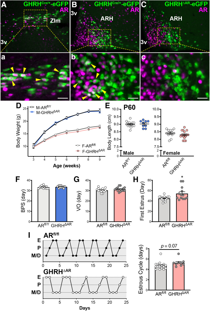
Mice with deletion of AR in GHRH cells have normal growth, but females show delayed pubertal completion. A, B, Representative fluorescent micrographs showing colocalization (arrowhead) of AR- (magenta) and GHRHCre/+-eGFP (green) immunoreactivity (-ir) in the ZIm (28.28 ± 9.93% of eGFP+ cells; A) and in the ARH (22.98 ± 3.30%; B) in males. C, Fluorescent micrograph showing AR-ir and GHRHCre/+-eGFP-ir in the ARH of GHRHΔAR mice. Note lack of colocalization (0.71 ± 0.81%), indicating successful deletion of AR in GHRH neurons. Higher-magnification micrographs of the selected area in A-C are shown in Aa, Bb, and Cc, respectively. D, Body weight progression in ARfl/Y (n = 10) and GHRHΔAR males (M; n = 16); and ARfl/fl (n = 17) and GHRHΔAR females (F; n = 21), by two-way repeated-measures ANOVA with Sidak's multiple comparisons. E, Body length at P60 in GHRHΔAR (n = 9) versus ARfl/Y (n = 14) males and GHRHΔAR (n = 16) versus ARfl/fl (n = 12) females. F, Day of BPS. G, Day of VO. H, First estrus. Note delay in pubertal completion (first estrus) in GHRHΔAR (n = 13) compared with controls (n = 15; t(15) = 2.55, p = 0.022). I, Representative estrous cycles of 2 mice from each genotype. No differences in estrous cycle were detected between genotypes (t(23) = 1.89, p = 0.071). *p < 0.05 (unpaired two-tailed Student's t test with Welch's correction). Each point represents 1 individual mouse. 3v, Third ventricle. Scale bar, 50 µm.

