Figure 7.
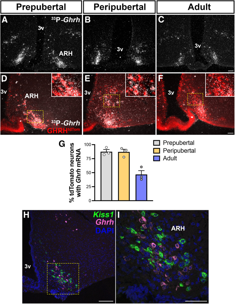
Changes in ARH Ghrh mRNA expression during development and localization with Kiss1 neurons. A–C, Representative darkfield micrographs showing Ghrh mRNA (hybridization signal) in the ARH of prepubertal, peripubertal, and adult (n = 3/group) females. D–F, Representative images of Ghrh mRNA (silver grains) distribution in ARH GHRHtdTom neurons in prepubertal, peripubertal, and adult females. Insets, Higher-magnification micrographs of the selected area. G, Quantification of ARH Ghrh mRNA coexpression with GHRHtdTom neurons during development. H, I, Representative confocal dual-fluorescent micrographs showing the distribution of ARH Kiss1 (green) and Ghrh (magenta) mRNA expression in diestrus phase. DMH, Dorsomedial nucleus of the hypothalamus; 3v, third ventricle. Each point represents 1 individual mouse. Scale bars: A–C, H, 100 µm; D–F, I, 50 µm.

