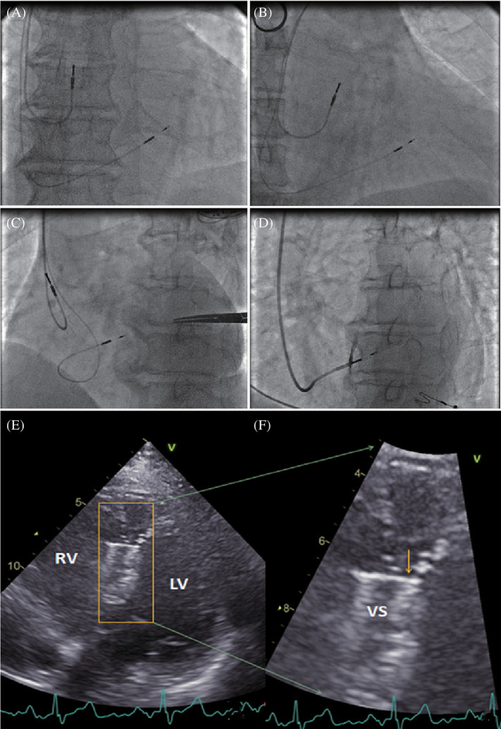
FIGURE 1.

LBBP lead location confirmed by fluoroscopy and post‐implant 2D echocardiography. Fluoroscopic imaging when the pacing lead was placed in the left bundle branch area which was beneath the left side of the ventricular septum in orthotopic view, the right anterior oblique 30° view and the left anterior oblique 40° view, A‐C. Contrast injection through the sheath in the left anterior oblique 35° view, D. F, Enlarged picture of, E, to show the pacing lead tip under the left endocardium of the septum. The arrow indicates the location of the pacing lead tip with a high density. LBBP, left bundle branch pacing; LV, left ventrium; RV, right ventrium; VS, ventricular septum
