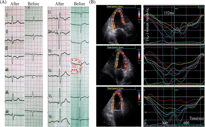
FIGURE 5.

An 86 years old male patient with left anterior fascicular block and I0 II0 AVB accepted LBBP using a 3830‐pacing lead. After LBB pacing, the left anterior fascicular block still existed (Figure 5A). 2‐D speckle‐tracking echocardiography was used to get the time‐systolic strain curve of the 18 segments after operation (Figure 5B); 2D‐TDmax of the 18‐segment systolic time to peak systolic strain was 152 ms (Figure 5B), which has no statistical difference compared with that of RVP group (148.62 ± 43.67). AVB, atrioventricular block; LBBP, left bundle branch pacing; RVP, right ventricular pacing
