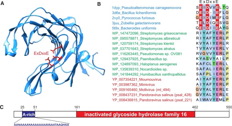
Figure 3.
MVP2 homologs are derived from inactivated GH16. (A) Structure of a representative GH16, kappa-carrageenase of Pseudoalteromonas carrageenovora (PDB ID: 1DYP; Michel et al. 2001). Acidic active site residues are shown using stick representation and are colored red. B. Alignment of representative GH16 (blue) as well as inactivated derivatives from Modules 1 to 3 (green) and viruses (red). Only the region harboring the active site (ExDxxE) of GH16 is shown. Active site triad is shown in white font on the red background. The full alignment can be found in Supplementary Fig. S2. (C) Domain organization of P. salinus MVP2 (psal_221). A-rich, alanine-rich region.

