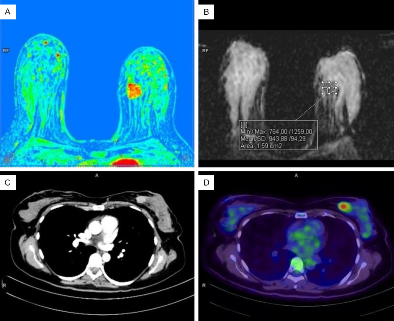
Figure 1.

Breast MRI (upper row) and whole-body PET/CT with 18F-FDG (lower row) in a patient with invasive breast cancer. A. The perfusion map shows a hypervascular tumor in the inner quadrants of the left breast. B. ADC map with an example of freehand ROI placement. C. CT scan with intravenous contrast (iopamidol) - a component of combined PET/CT. D. A significant increase in tumor metabolism was detected via PET/CT with 18F-FDG.
