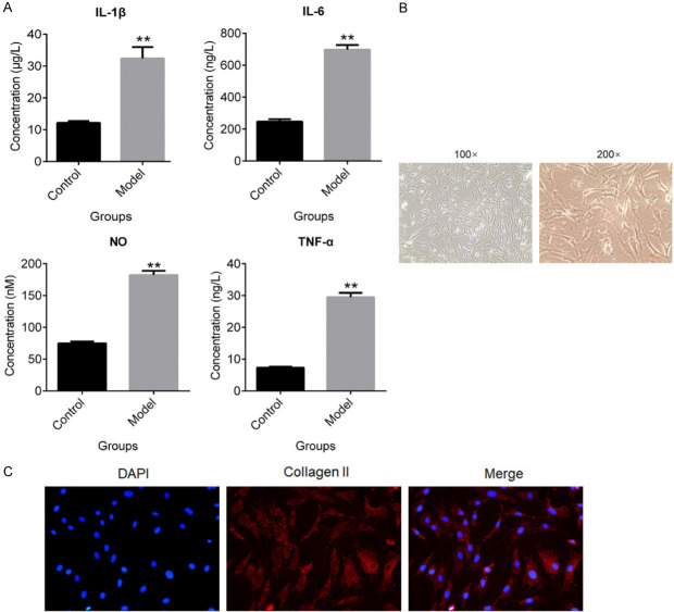
Figure 1.
OA was established on rats and the chondrocytes were isolated. A. The concentration of IL-1β, IL-6, NO and TNF-α was evaluated by ELISA (**P < 0.01, vs. Control). B. The morphology of chondrocytes was pictured by microscope. C. The expression level of collagen II was determined by immunofluorescence assay.

