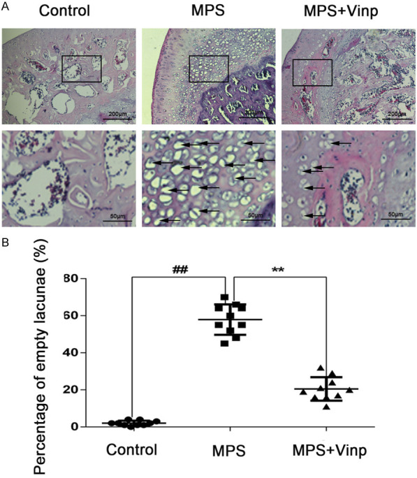
Figure 5.

Morphological changes in the femoral head as seen on H&E staining in the control, MPS, and MPS+Vinp groups. In the femoral head of control group, no empty bone lacunae could be found (A). Massive empty lacunae (black arrows) surrounded by necrotic marrow cells are seen in the MPS group, with fewer empty bone lacunae in the MPS+Vinp group. The ratio of empty lacunae was visibly higher in the model group than in either the control or Vinp group (B). ##P < 0.01 compared with Control group. **P < 0.01 compared with MPS group.
