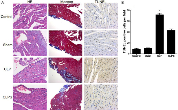
Figure 7.
Monocyte mobilized to myocardial tissue in sepsis and led to myocardial damage. A: The first column: representative HE staining showed that monocyte accumulated to myocardial tissue in CLP group. No obviously inflammatory cells occurred in control, sham and CLPS. Myocardial dissolving and inflammatory cell infiltration was observed in sepsis group (400*). The second column: representative Masson staining photograph. Collagen fiber was detected in sepsis instead of muscle fibers (100*). The third column: representative TUNEL staining showed that apoptotic cardiomyocytes were no obviously changed between control and sham (400*). Markedly increased apoptotic cardiomyocytes in the CLP group. In CLPS, the number of apoptotic cardiomyocytes is lower than CLP. B: Histogram showed the number of apoptotic cardiomyocytes among the four groups (n=5 for control and sham, n=10 for CLP and CLPS, *P<0.05 vs control, sham and CLPS).

