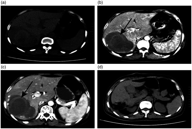
Figure 1.
a. On admission, chest CT scan revealed a mass flaky shadow of the right liver lobe. b. The first abdominal augmentation CT examination showed that the right liver lobe had a circular mixed density mass of about 10 × 9 cm, which had a clear boundary and contained a ring-shaped, strip-like slightly high-density shadow (shown by a white arrow). c. After 2 weeks, the high-density hemorrhage foci in the lesion increased compared with 2 weeks before. d. A postoperative CT scan showed a small amount of effusion.
CT, computed tomography.

