
Fig. 1.
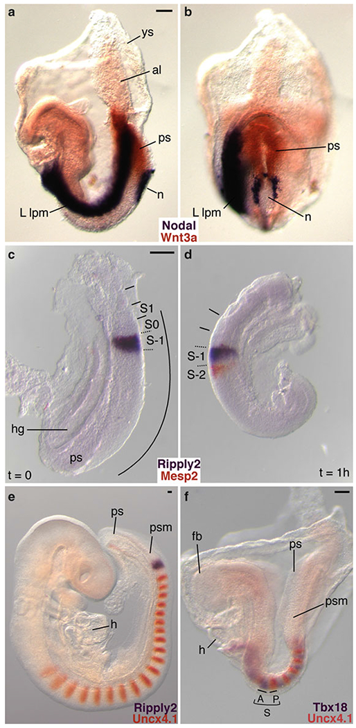
Examples of two-color WISH performed on mouse embryos and cultured embryo explants. In all cases, DIG-labeled RNA probes were detected with BM purple, and FLU-labeled RNA probes were visualized with INT/BCIP. (a, b) Two-color WISH analysis of a 4 somite stage (E8.2) embryo demonstrating the complementary expression of genes encoding the secreted signaling molecules Nodal (purple) and Wnt3a (orange). Nodal is asymmetrically expressed in the node periphery and in the (left) lateral plate mesoderm [9,10], while Wnt3a mRNA is restricted to the midline primitive streak [11] (Reproduced from ref. 5 with permission from Development). (c, d) Embryo-half culture experiments illustrate how dynamic, oscillating gene expression patterns can be visualized with two-color WISH. The bHLH transcription factor, Mesp2 (orange), and the transcriptional corepressor Ripply2 (purple), are important for segment boundary formation [4, 5,12,13]. In the uncultured half-embryo explant (t = 0), both genes are coexpressed in S-1 in the anterior presomitic mesoderm (c). After culturing the complementary half-embryo explant for 1 h, Ripply2 expression remains in S-1 while Mesp2 expression is activated in S-2 (d). (e) Analysis of E9.5 embryos shows that the paired type homeodomain transcription factor Uncx4,1 (orange) is expressed in the caudal half of segmented somites [14–16], and in the posterior half of SO immediately adjacent to the broad somite-wide stripe of Ripply2 expression (purple) in S-1 [4]. (f) Two-color WISH clearly illustrates the complementary expression patterns of segment polarity markers. Expression of the anterior half-somite marker Tbx18 (purple) complements Uncx4,1 expression in the caudal half [5, 17]. All embryo views are lateral with the exception of (b), which is ventroposterior. ys yolk sac, al allantois, ps primitive streak, n node, L lpm left lateral plate mesoderm, hg hindgut, S somite, S-1 presumptive somite, SO forming somite, S1 first newly formed somite, curved line or psm presomitic mesoderm, h heart, fb forebrain, A anterior half somite, Pposterior half somite. Scale bars: 100 μm