
Fig. 2.
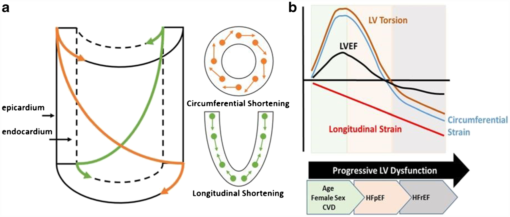
Twist mechanics and altered strain contributions to left ventricular ejection fraction with age, sex, and cardiovascular disease. a Left ventricular myofiber architecture, changing from a left-handed helix in the subepicardium to a right-handed helix in the subendocardium. Contraction of these two opposing myofiber layers gives rise to circumferential and longitudinal shortening about the long axis of the cylinder. Note the longer lever arm of the subepicardial fibers compared with the subendocardial fibers. When both layers contract simultaneously, the epicardial fibers have a mechanical advantage, dominating the overall direction and magnitude of rotation. This mechanical advantage is augmented in conditions with impaired subendocardial function and/or a greater subepicardial radius (i.e. concentric hypertrophy). b Conceptual model illustrating patterns of change in left ventricular tissue deformation, twist mechanics, and ejection fraction through the onset of early mechanical dysfunction (associated with age, sex, and cardiovascular comorbidities), heart failure with preserved ejection fraction (HFpEF), and heart failure with reduced ejection fraction (HFrEF)