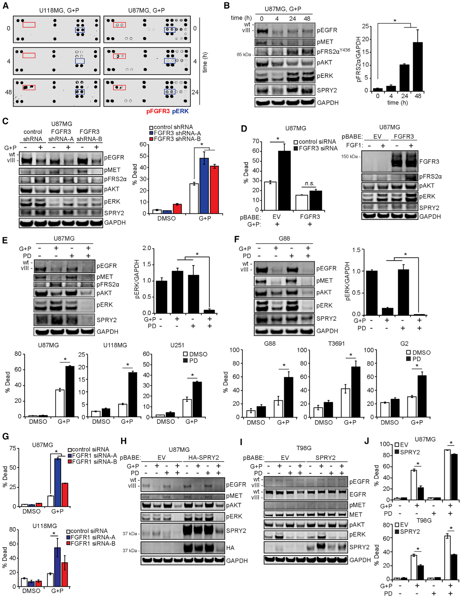
Figure 3. FGF Receptors Drive ERK Reactivation and Resistance to EGFR and MET Inhibitors.

(A) U87MG and U118MG cells were treated with 10 μM gefitinib (G) + 3 μM PHA665752 (P) for the indicated times. Lysates were prepared and incubated with antibody microarrays, with signals for U87MG quantified by densitometry in Figure S3A.
(B) The U87MG lysates from (A) were analyzed by western blotting using antibodies against the indicated proteins, with the pFRS2α signal quantified by densitometry and normalized to values at t = 0 h.
(C) U87MG cells expressing control or FGFR3-targeting shRNA were treated with 10 μM G + 3 μM P or DMSO for 24 h. Cell lysates were analyzed by western blotting using antibodies against the indicated proteins. Cells treated in parallel for 48 h were analyzed by flow cytometry for cell death.
(D) U87MG cells transduced with a vector encoding an RNAi-resistant FGFR3 or an empty vector (EV) were transfected with control or FGFR3-targeting siRNA for 48 h. Cells were then treated for 24 h with 10 μM G + 3 μM P, and cell death was measured by flow cytometry. Cell death data for DMSO-treated cells are shown in Figure S3E. In parallel, untransfected cells were treated with 50 ng/mL FGF1 for 5 min, and cell lysates were analyzed by western blotting using antibodies against the indicated proteins.
(E) U87MG cells were treated for 24 h with 10 μM G + 3 μM P, 0.5 μM PD173074 (PD), G + P + PD, or DMSO. Lysates were analyzed by western blotting using antibodies against indicated proteins, with the pERK signal quantified by densitometry and normalized to DMSO-treated values. U87MG, U118MG, and U251 cells were treated with the same inhibitors for 48 h, and cell death was measured by flow cytometry.
(F) G88 cells were treated for 48 h with 5 μM G + 1 μM P, 0.5 μM PD, G + P + PD, or DMSO before lysis and western blot analysis. G88, T3691, and G2 cells were treated with the same inhibitors for ≤72 h, and cell death was measured by flow cytometry.
(G) U87MG and U118MG cells were transfected with control or FGFR1-targeting siRNA for 48 h and then treated for 24 h with 10 μM G + 3 μM P, or DMSO, and cell death was measured by flow cytometry.
(H and I) U87MG cells (H) or T98G cells (I) were transduced with vectors encoding SPRY2 or an EV and treated for 24 h with 10 μM G + 3 μM P, 0.5 μM PD, G + P + PD, or DMSO. Lysates were analyzed by western blotting using antibodies against indicated proteins. Hemagglutinin (HA)-tagged SPRY2 was used in U87MG cells to differentiate abundant endogenous SPRY2 from ectopic SPRY2.
(J) SPRY2-overexpressing U87MG or T98G cells were treated with the same inhibitors for 48 h, and cell death was measured by flow cytometry. Throughout the figure panels, representative blot or array images are shown, and error bars indicate means ± SEMs for three replicates; *p < 0.05 for the indicated comparisons.
