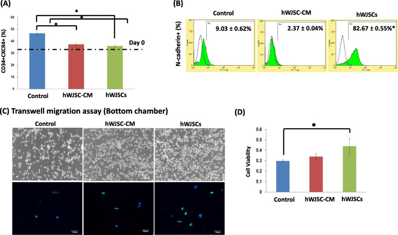
Fig. 5.
Cell adhesion markers and migration of UCB CD34+ cells expanded with hWJSCs and hWJSC-CM. A Note the percentage of the most primitive CD34+CXCR4+ cells in the hWJSCs and hWJSC-CM-expanded groups was closest to the initial seeding cell numbers on day 0 (dotted line) compared to the controls. B Note significantly greater percentage of N-cadherin+ cells after expansion of CD34+ cells with hWJSCs as compared to controls. C (a–c) Phase-contrast images of the bottom chamber of transwell migration assays. Note increased number of CD34+ cells that have migrated from the top to the bottom chamber when cultured in the presence of hWJSCs and hWJSC-CM compared to controls. (d–f) There was an increased number of N-cadherin positive cells among the migrated cells expanded with hWJSCs and hWJSC-CM compared to controls. D Note significantly greater viable CD34+ cell numbers (MTS assay) lying in the bottom chamber of transwell migration chambers after expansion with hWJSCs as compared to controls. All values are expressed as mean ± SEM of 3 biological samples with 3 replicates for each sample. *p < 0.05

