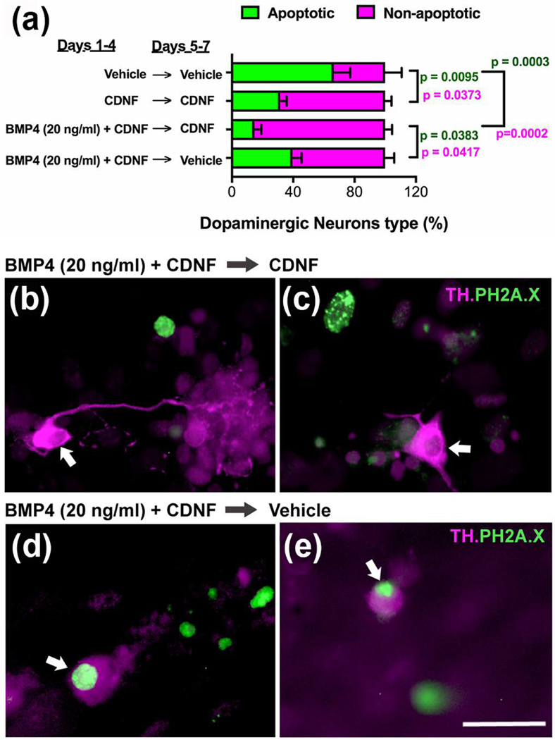
Figure 11. Exposure to BMP4 enhances the development of dependence of dopaminergic neurons on CDNF for survival.
a. Isolated ENCDC were cultured for 7 days. As in Fig. 4, the full culture time was divided into an initial period of 4 days and a subsequent period of 3 days during which the cells were either maintained in vehicle or in CDNF (100 ng/ml). Cells were exposed during the initial period to vehicle, CDNF (100 ng/ml; n = 4 replicates), or to 20 ng/ml BMP4 plus CDNF (5 replicates) in order to evaluate the effects of CDNF withdrawal. Expression of TH immunoreactivity was used to identify dopaminergic neurons and that of PH2A.X was employed to identify cells undergoing apoptosis. The proportion of the population of dopaminergic neurons that was apoptotic (green) or non-apoptotic (magenta) was quantified for each of the tested conditions. A significantly smaller proportion of dopaminergic neurons was apoptotic in cultures continuously exposed to CDNF than in cultures exposed continuously to vehicle. Initial exposure of cultures to BMP4 (20 ng/ml) resulted in a significant further reduction of the proportion of apoptotic dopaminergic neurons as long as CDNF was maintained during the subsequent 3 days in vitro; however, a significant increase in the proportion of dopaminergic neurons that was apoptotic occurred when CDNF was withdrawn and cells were exposed only to vehicle for the final 3 days in culture [live dopaminergic as %total dopaminergic, one way Anova F(4, 19) = 5.435 p = 0.0043; apoptotic dopaminergic as % total dopaminergic, one way Anova F(3, 14) = 10.6 p=0.0007]. b, c. Illustration of cultures initially exposed to BMP4 (20 ng/ml) plus CDNF and then maintained in the presence of CDNF. Note that although some cells in the cultures have PH2A.X-immunoreactive nuclei and thus are undergoing apoptosis, the dopaminergic neurons in the field (arrows) are healthy and exhibit abundant neurite outgrowth. d. e. Illustration of cultures initially exposed to BMP4 (20 ng/ml) plus CDNF but then subjected to CDNF withdrawal. Note that dopaminergic neurons now have PH2A.X-immunoreactive nuclei and thus are undergoing apoptosis; the apoptotic dopaminergic neurons (arrows) do not exhibit abundant neurite outgrowth. The bar = 35 μm.

