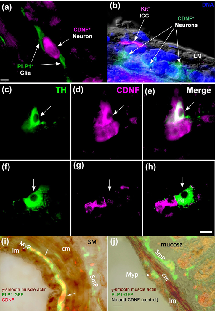
Figure 2. Enteric CDNF immunoreactivity is found in subsets of dopaminergic and other neurons but not in enteric glia, interstitial cells of Cajal, or smooth muscle.
a. Section of bowel wall cut through the myenteric plexus of a PLP-1-GFP mouse immunostained to demonstrate CDNF. PLP-1 marks enteric glia. GFP fluorescent processes (green) of enteric glial cells closely interact with a CDNF-immunoreactive neuron (magenta). Note absence of CDNF immunoreactivity from glia. The bar = 10 μm (a, b). b. Bowel wall cut through the myenteric plexus of a Cdnf +/+ mouse immunostained to demonstrate immunoreactivities of Kit (magenta) and CDNF (green). Kit marks ICC. DNA was demonstrated with bisbenzimide (blue) and the field was also viewed in differential interference contrast. The plexus of ICC that surround myenteric ganglia lack CDNF immunoreactivity. No CDNF immunoreactivity can be seen in the longitudinal muscle (LM) layer outside of the ganglion. c-h. Whole mounts of submucosa containing ganglia immunostained for TH (green) to mark dopaminergic neurons and CDNF (magenta). c-e. The illustrated ganglion contains a TH-immunoreactive neuron that displays coincident CDNF immunoreactivity. Note in D and E that not all CDNF-immunoreactive neurons are TH-immunoreactive. The arrow locates a dopaminergic neuron in each panel. f-i. The illustrated ganglion contains a TH-immunoreactive neuron that is not CDNF-immunoreactive. Note in g and h that several CDNF-immunoreactive neurons lack TH-immunoreactivity. The arrow shows the location of the dopaminergic neuron in each panel. The bar = 16 μm (c-h). i-j. Enteric glia in PLP-1-EGFP mice (bowel sections). Smooth muscle was immunostained with antibodies to γ–smooth muscle actin and CDNF. In a control section, antibodies to CDNF were omitted. (i) Animal at about 2 months of age (P58). γ–smooth muscle actin antibodies directly coupled to horseradish peroxidase (HRP) label smooth muscle cells (brown). Triple labeling demonstrates CDNF (red fluorescence) and GFP (glia; green fluorescence). HRP reaction product photographed in white light and merged with the fluorescent images of EGFP and CDNF. Myenteric plexus = MyP, submucosal plexus = SmP, and submucosa = SM. Arrows locate CDNF-immunoreactive neurons. Antibodies to CDNF fail to label smooth longitudinal (lm) or circular (cm) muscle or glia. (j) Control. Antibodies to CDNF were omitted. Section is PLP-1-EGFP bowel from P21 mouse. Mucosa is indicated. Bar (i and j) = 16 μm.

