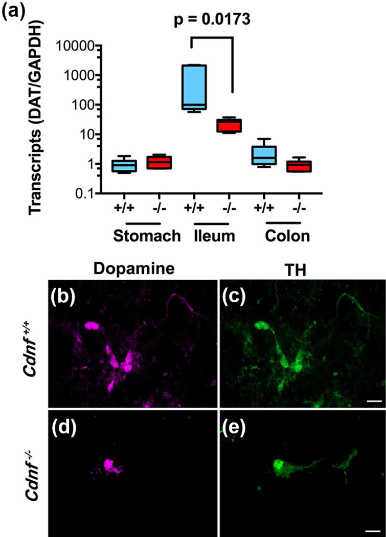
Figure 3. Markers of dopaminergic neuronal development are less abundant in the ileum of Cdnf −/− mice than in Cdnf +/+ animals.
a. Transcripts encoding DAT were employed as a marker for the development of dopaminergic neurons in the murine stomach, ileum, and colon at P7. Transcripts encoding DAT were far more abundant in the ileum than in either of the other regions of the bowel (note that the ordinate is logarithmic) [one way Anova, F(5, 30) = 3.088 p = 0.0230]. Transcripts encoding DAT in the ileum were significantly more abundant in the Cdnf +/+ mice (blue) than in Cdnf −/− animals (red); [Sidak’s multiple comparison test p = 0.0173]. Each bar represents the mean values derived from analyses of 6 samples pooled from 2 animals of each genotype. b. Visualization of dopamine and TH immunoreactivities in the submucosal plexus of the murine ileum (9-month-old). In both Cdnf +/+ and Cdnf −/− mice, these markers are fully coincident; however, dopamine- and TH-immunoreactive cells are more numerous in Cdnf +/+ than in Cdnf −/− mice. The bar = 32 μm.

