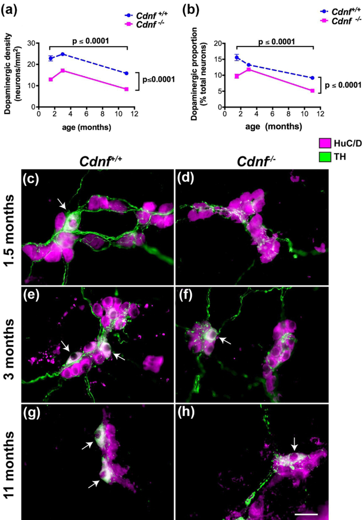
Figure 4. Both the packing density and the proportion of dopaminergic neurons are decreased as a function of age in Cdnf −/− compared to Cdnf +/+ mice.
a. Packing density (number/unit area) of dopaminergic neurons is plotted as a function of age in Cdnf +/+ (blue) and Cdnf −/− mice (magenta). Data were obtained from 60–102 measurements from 3–6 mice at each age and genotype. The decline from 1.5-month-old to 11-month-old is significant in mice of both genotypes; however, at each age examined the density is significantly greater in Cdnf +/+ than in Cdnf −/− animals [two way Anova F(2, 476) = 73.63]. Coincident immunoreactivity of TH and HuC/D was used to identify dopaminergic neurons. b. The proportion of total neurons (HuC/D-immunoreactive) that were dopaminergic (TH-immunoreactive) is plotted as a function of age. The proportion of dopaminergic neurons declines significantly as a function of age in both Cdnf +/+ and Cdnf −/− mice; moreover, the proportion of dopaminergic neurons is always greater in Cdnf +/+ than in Cdnf −/− animals [two way Anova F(2, 403) = 61.62]. c-h. Representative images of TH (green) and HuC/D immunoreactivities (magenta) in the ileal submucosal plexus of Cdnf +/+ (c, e, g) and Cdnf −/− mice (d, f, h) at 1.5 (c, d), 3 (e, f), and 11 months of age (g, h). The bar = 35 μm.

