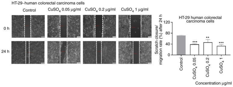
Figure 7.
The impact of CuSO4 (0.05, 0.2 and 1 µg/ml) on HT-29 human colorectal carcinoma cells migratory capacity following 24 h of stimulation. Scratch widths were initially recorded by bright field microscopy, 0 and 24 h post exposure. Scale bars represent 100 µm. The results are expressed as scratch closure/migration rate (%) following 24 h of stimulation. The data represent the mean values ± SD of three independent experiments performed in triplicate. One-way ANOVA analysis was applied to determine the statistical differences from the control cells, followed by Dunnett's multiple comparisons post-test (**P<0.01, ***P<0.001).

