Figure 3.
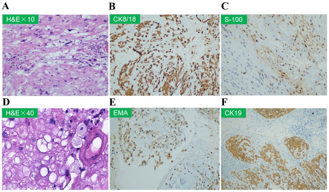
(A-F) Sacral needle core biopsy of the T11 vertebra. (A and D) Representative images of H&E-stained sections (magnification, x10 and x40, respectively) showing cytoplasmic vacuolation. Representative images of immunohistochemical staining for (B) CK8/18, (C) S-100, (E) EMA and (F) CK19. H&E, hematoxylin and eosin; CK, cytokeratin; EMA, epithelial membrane antigen.

