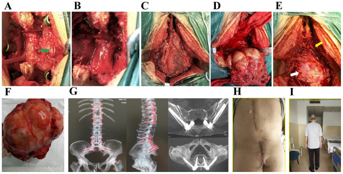
Figure 4.
(A and B) Stage I resection of the thoracic intraspinal mass protruding into the spinal canal (arrow) after posterior decompression. (C and D) Stage II extended (C) sacral tumor resection with (D) removal of S1 and S2 spaces of the upper boundary of the tumor. (E) The S1 nerve (yellow arrow) and the posterior peritoneum of the rectum (white arrow) remained intact after surgery. (F) The resected tumor measured ~14x15 cm. (G) Computed tomography with maximum intensity projection image reconstruction after posterior thoracic decompression with screw-rod internal fixation and sacral tumor resection. (H) Inspection of incision healing after surgery. (I) Walking ability was restored 2 weeks after surgery.

