
Figure 2.
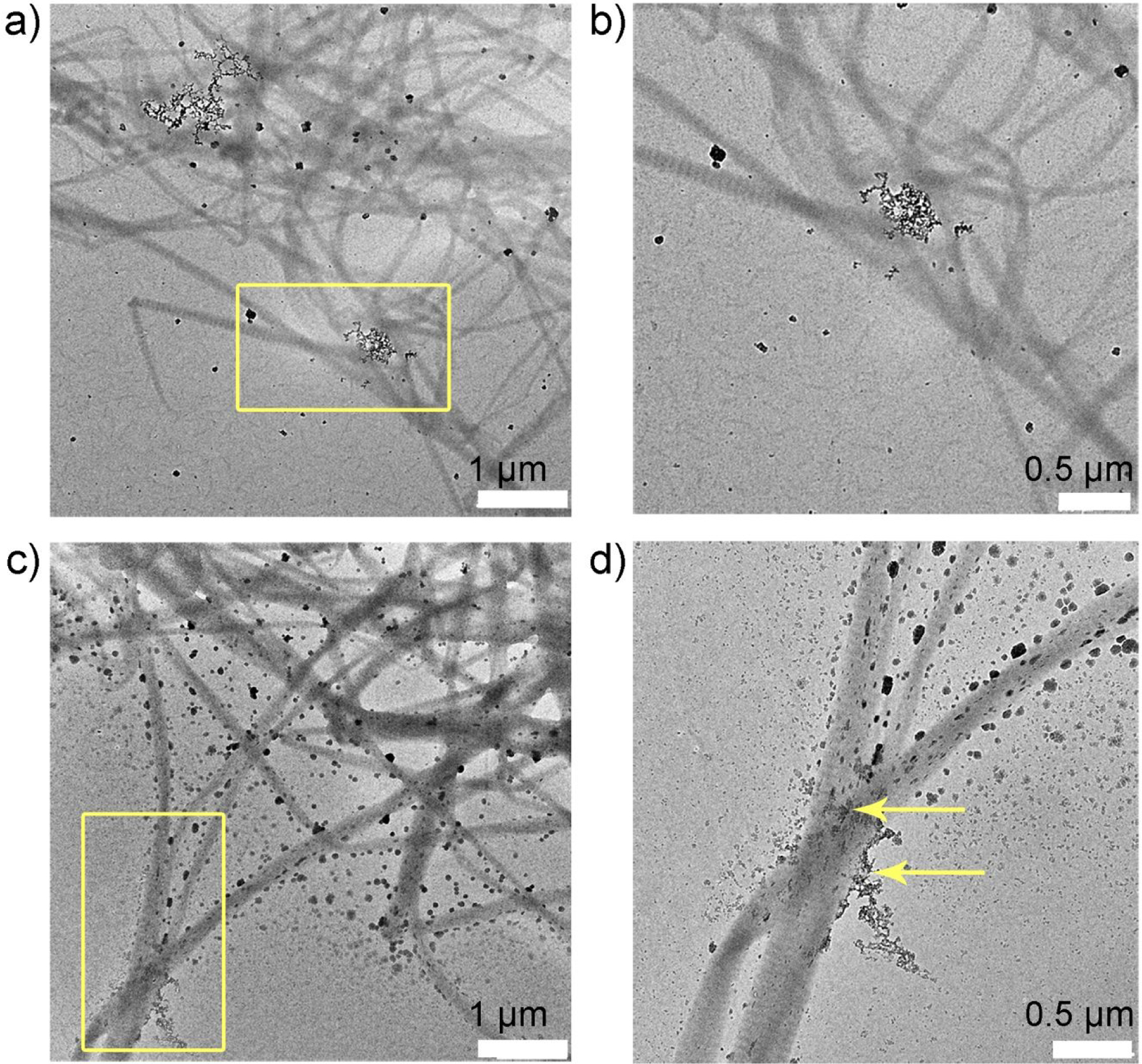
Collagen-P26 mineralization after 1 hour. Unstained TEM images of collagen mineralization in vitro for 1 h in the control sample (no P26) at (a) low and its corresponding (b) high resolution of yellow square in (a). There are very few amorphous particles seen on the grid surface. TEM images of collagen mineralization in vitro for 1 h in the presence of P26 at (c) low and its corresponding (d) high resolution of yellow square in (c). Some of the fibrils in grid space (d) appear swollen with the precipitation of amorphous minerals on the surface (yellow arrows).