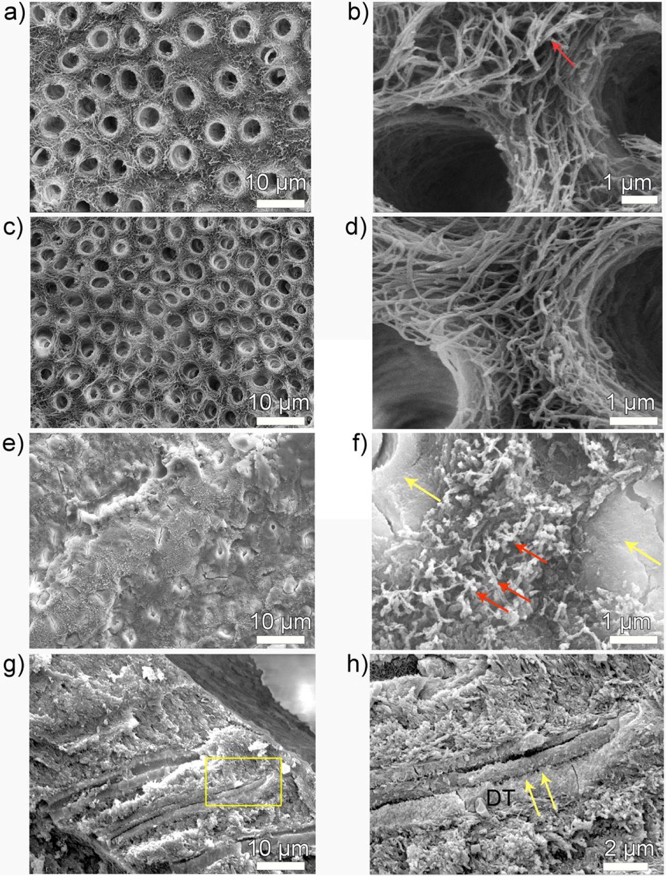
Figure 4.

SEM images of demineralized dentin surface before and after remineralization. (a, b) 3-day demineralized dentin, (c, d) control and (e-h) P26-treated dentin discs after incubation in artificial saliva in pH 7.0 at 37 °C. Images (b, d, f, h) are the high resolution corresponding magnified images of (a, c, e and g) respectively. (b) is a high-resolution SEM image of demineralized, mineral-depleted, collagen fibrils of intertubular dentin, giving a smooth ribbon-like appearance (red arrow). (c, d) There is no HAP crystallization on the surface of dentin in the control dentin discs and the tubules remain open. (e, f). After P26 application, the surface of dentinal tubule is completely occluded by a dense layer of HAP mineral precipitate (yellow arrows) and the exposed collagen fibrils are embedded in electron-dense mineralized areas (red arrows in f). (g, h) Cross-sections of the dentin discs show penetration of mineral ions up to 15 μm into the subsurface adhering firmly to the walls of the dentinal tubules (yellow arrows). DT- dentinal tubule.
