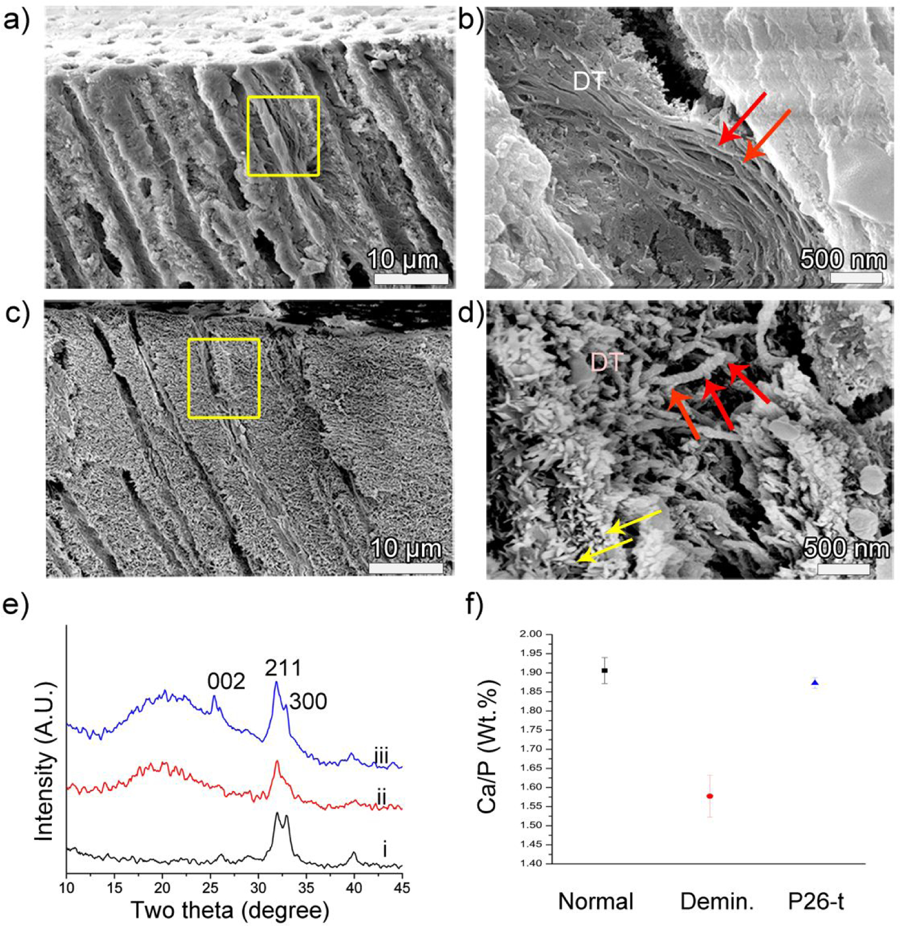
Figure 5.

Cross-sectional SEM images of dentin after remineralization. (a, b) control and (c, d) P26-treated dentin discs after 10 days of incubation in artificial saliva in pH 7.0 at 37 °C. Images (b, d) are the corresponding magnified images showing a single dentinal tubule of (a, c) in the yellow squares respectively. Figure (b) depicts the lack of mineralization of collagen fibers inside a dentinal tubule in the control. (d) After P26 application there is a marked depth of mineral deposition inside the tubule up to 15 μm from the surface. Newly formed crystals can be seen growing along the walls of the tubules (yellow arrows) and the diameter of the individual collagen fibers increases with time, giving a ‘beaded string-like’ appearance due to HAP precipitation (red arrows). (e) XRD spectra of (i) sound dentin, (ii) demineralized and (iii) remineralized dentin surfaces after P26 application. Distinct peaks for peptide-treated dentin discs indicate improved crystallinity of HAP when compared to ii. (f) EDX data showing Ca/P weight ratio (weight %) on the surfaces of healthy, demineralized and P26-treated dentin (n=3).
