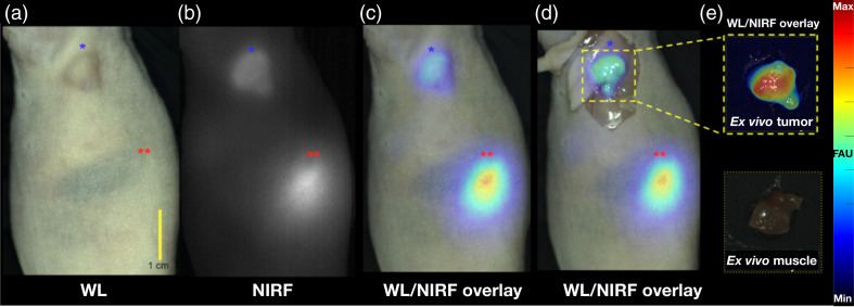
Fig. 3.
High in vivo contrast detection of fluorescently-labeled subcutaneous tumors under ambient light using simultaneous WL and NIRF image acquisition on the OnLume imager. Representative images of a HCT116-SSTR2 tumor imaged in situ 48 h after the injection of -MMC(IR800)-TOC under (a) WL, (b) NIRF, (c) WL with NIRF overlay, and (d) WL with NIRF overlay with skin retracted. (e) WL with NIRF overlay of excised tumor and muscle. The tumor is labeled by the blue star (*), and the kidney is labeled with two red stars (**). Fluorescence arbitrary units (FAU).

