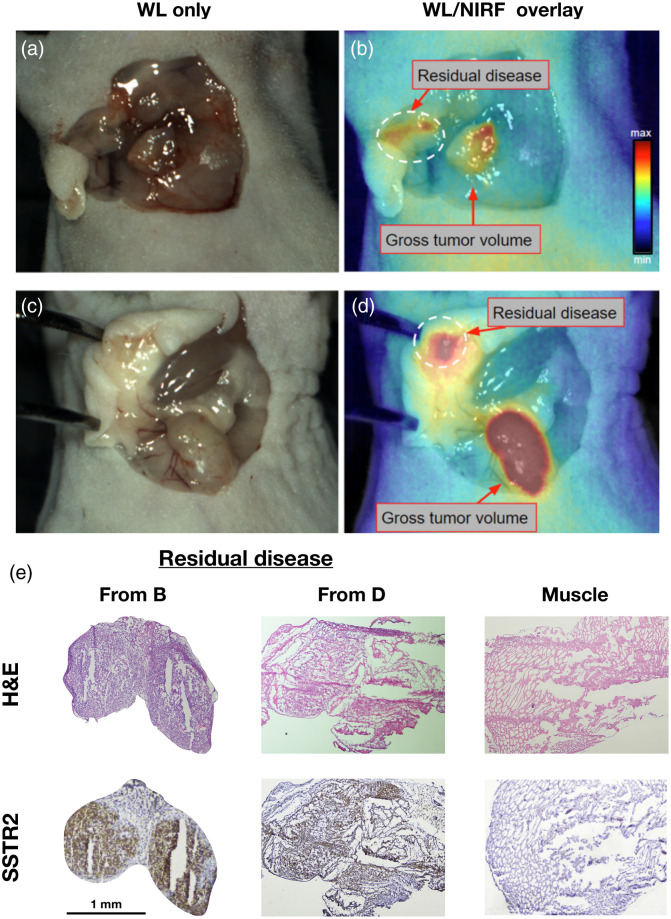
Fig. 7.
Real-time, intraoperative detection of residual disease using FGS under ambient light. (a) and (c) WL images of the wound bed after tumors were resected using visual inspection and palpation. (b) and (d) Corresponding NIRF images acquired with the OnLume imager revealed residual fluorescence that was not visible with the naked eye (dashed circle). (e) Histological analysis of these microlesions confirmed for cancer positivity (H&E) and SSTR2-expression (IHC). Muscle staining was performed as a negative control. Reproduced with permission from Ref. 34 (Video 1, MP4, 50 MB [URL: https://doi.org/10.1117/1.JBO.25.12.126002.1]) (Video 2, MP4, 47 MB [URL: https://doi.org/10.1117/1.JBO.25.12.126002.2]).

