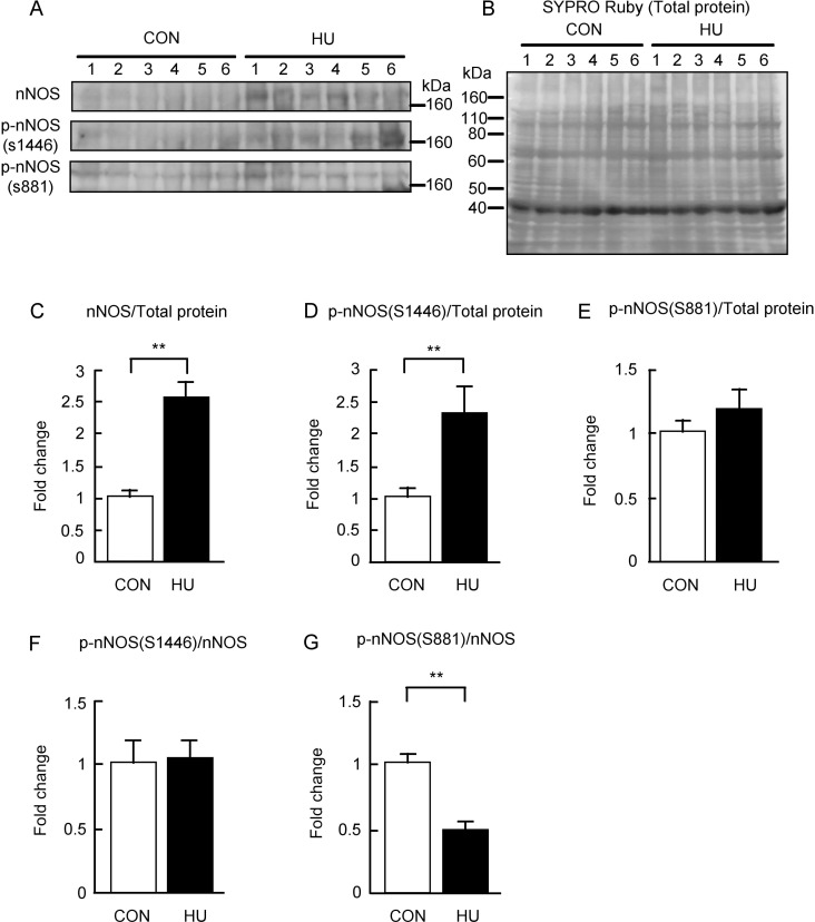
Fig 2. Changes in the expression levels of nNOS and phosphorylated nNOS after hindlimb unloading.
(A) Western blots showing immunoreactivities of nNOS, phosphorylated nNOS at S1446, and phosphorylated nNOS at S881. (B) Total membrane protein detected by SYPRO Ruby staining. (C-E) Comparisons of nNOS, phosphorylated nNOS at S1446, and phosphorylated nNOS at S881 normalized to total protein between groups are indicated in C, D, and E, respectively. (F, G) Comparisons of phosphorylated nNOS at S1446 and phosphorylated nNOS at S881 normalized to nNOS expression levels between groups are indicated in F and G, respectively. CON, control. HU, hindlimb unloading. Data are expressed as means ± SE. **significantly different from CON (p < 0.01).

