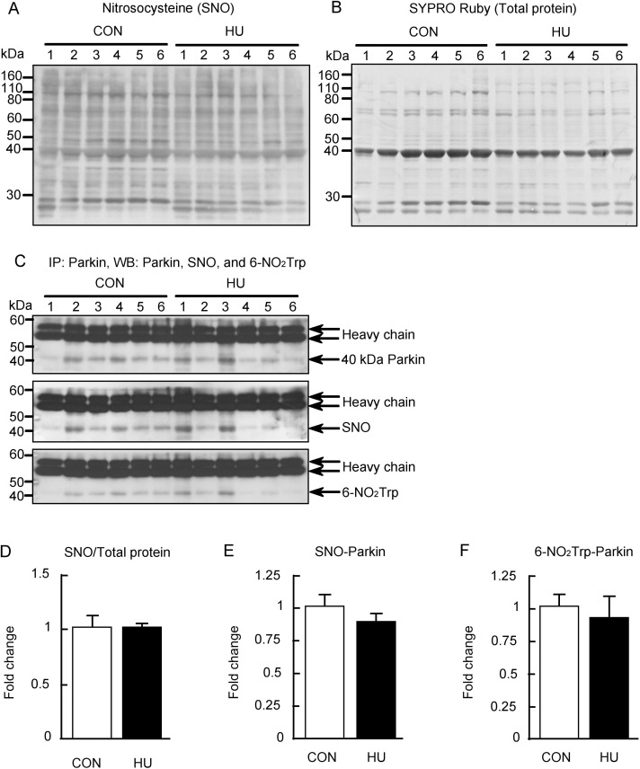
Fig 5. S-nitrosylation and tryptophan nitration of Parkin after hindlimb unloading.
(A) Nitrosocysteine (SNO) detected by western blot analysis. (B) Total membrane protein detected by SYPRO Ruby staining. (C) Immunoprecipitation performed using anti-Parkin antibody, and Parkin, SNO, and 6-nitrotryptophan (6-NO2Trp) immunoreactivities were detected by western blot analysis. (D) Comparisons of SNO immunoreactivity normalized to total protein between groups. (E, F) Comparisons of S-nitrosylated (SNO)-Parkin and tryptophan nitrated (6-NO2Trp)-Parkin normalized to Parkin expression levels between groups are indicated in E and F, respectively. CON, control. HU, hindlimb unloading. Data are expressed as means ± SE.

