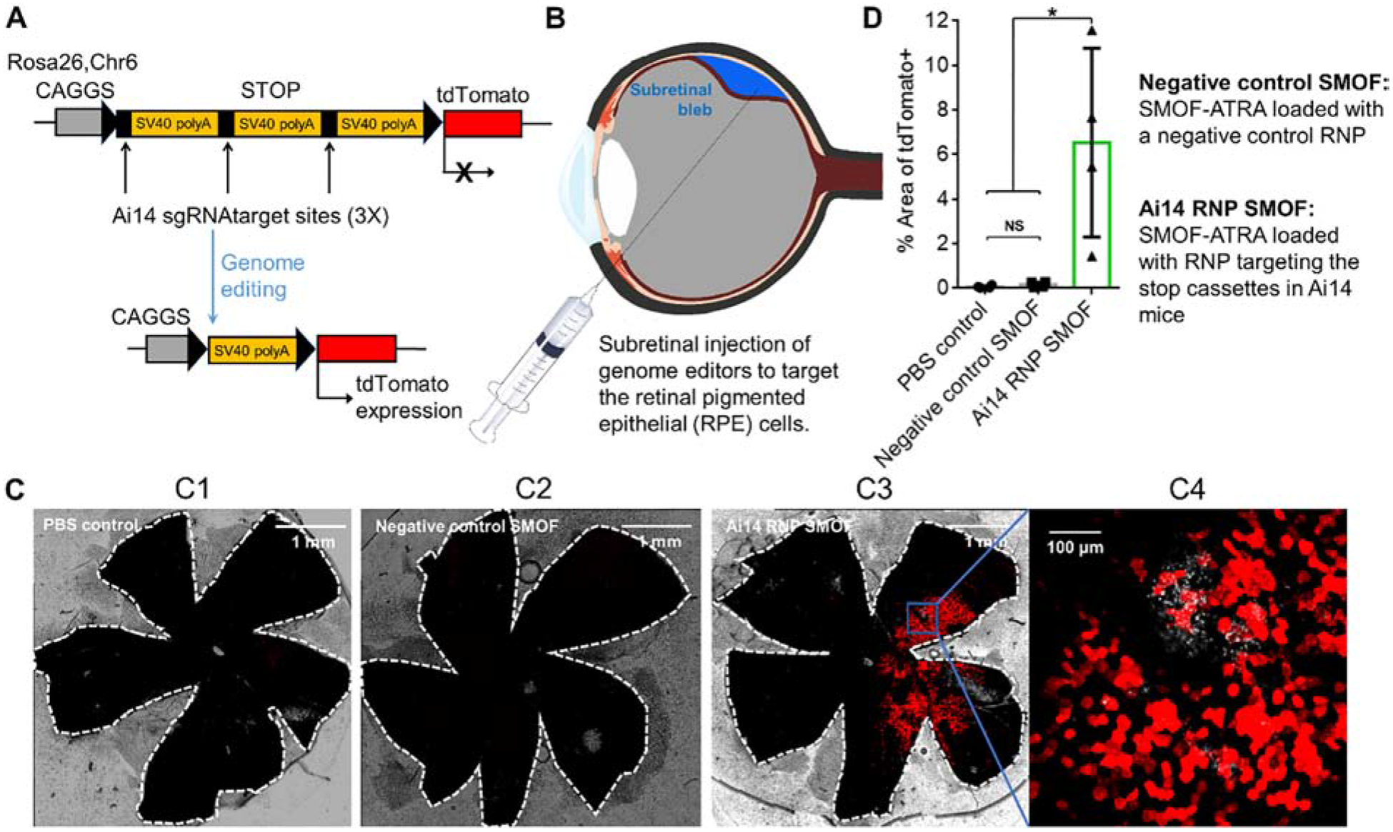
Figure 7. SMOF NPs induced efficient genome editing in vivo in Ai14 mice via local administration.

(A) The tdTomato locus in the Ai14 reporter mouse. A stop cassette containing 3 Ai14 sgRNA target sites prevents downstream tdTomato expression. RNP guided excision of the stop cassette results in tdTomato expression. (B) Illustration of SMOF NP subretinal injection targeting the RPE tissue. (C) Representative images of tdTomato+ signal (red) 13 or 14 days after subretinal SMOF-ATRA injection. The whole RPE layer was outlined with a white dotted line. C1: RPE floret of Ai14 mouse subretinally injected with PBS. C2: RPE floret of Ai14 mouse subretinally injected with negative control SMOF-ATRA (SMOF-ATRA encapsulating RNP with negative control sgRNA). C3: RPE floret of Ai14 mouse eye subretinally injected with SMOF-ATRA encapsulating RNP targeting the Ai14 stop cassette (i.e., Ai14 RNP SMOF), and C4: zoom-in image (20X magnification) of genome-edited RPE tissue induced by RNP-loaded SMOF-ATRA. (D) Genome editing efficiency as quantified by percent of the area of whole RPE tissue with tdTomato+ signals. NS: not significant, *: p<0.05.
