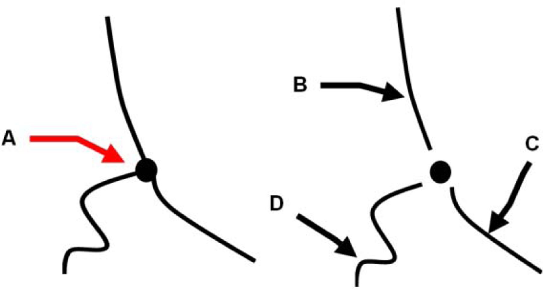
Figure 2:

Schematic for the definition of select morphological features. The simplified tumor angiogenic network from centerlines of the tubular structures contain vessels with branching points or nodes (A). Individual vessel segments, edges are counted after the removal of the branching point. (B), (C), and (D) denote individual vessels with gradually increased tortuosity and different vessel length.
