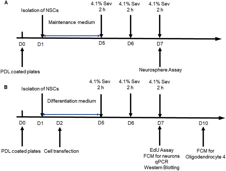
Fig 1. Diagrams for the experimental design.
A. NSCs were cultured in DMEM-F12 (1:1) maintenance medium containing 20 ng/mL of hEGF, bFGF, 1% N2 and 2% B27 for four days. The NSCs were exposed to sevoflurane (4.1% sevoflurane in 60% O2) or control 60% O2 alone 2 h daily for three consecutive days. The self-renewal of NSCs was analyzed by neurosphere formation. B. The NSCs (6 x 10⁵ cells/well) were cultured onto coverslips that had been coated with poly-l-lysine/laminin in DMEM-F12 (1:1) differentiation medium containing half concentration of growth factors to induce their differentiation for four days. The NSCs were exposed to sevoflurane (4.1% sevoflurane in 60% O2) or control 60% O2 alone 2 h daily for three consecutive days. The differential neurons and oligodendrocytes and astrocytes were characterized at day 7 and 10 post induction, respectively.

