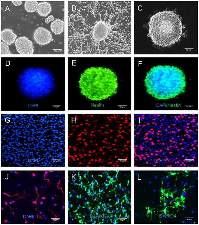
Fig 2. NSCs have the potential of self-renewal and multidirectional differentiation.
NSCs were culture for neurosphere formation and differentiation, as described in the method section. The formed neurospheres were imaged and the differentiated neurons, astrocytes and oligodendrocytes were characterized by indirect immunofluorescence. Data are representative images of different types of cells from three separate experiments. A. A microscopic image (magnification 10 x) of typical neurospheres. Scale bar = 100 um. B. The morphology of differentiated cells from NSCs. C. Light microscopy viewed NSCs. Magnification 20 x, Scale bar = 50 um. D-F. Indirect immunofluorescent analysis of neurosphere using anti-Nestin and Alexa Fluor 488-conjugated donkey anti-mouse IgG as well as DAPI. Magnification 20 x, Scale bar = 50 um. G-I. EdU+ cells indicate cell proliferation. Magnification 20 x, Scale bar = 50 um. J-L. Indirect immunofuorescent analysis of differentiated Tuj1+ neurons, GFAP+ astrocytes and O4+ oligodendrocytes using anti- Tuj1, anti-GFAP, anti-O4 and Alexa Fluor 574-labeled donkey anti-rabbit and Alexa Fluor 488-conjugated donkey anti-mouse as well as DAPI, respectively. Magnification 20 x, Scale bar = 50 um.

