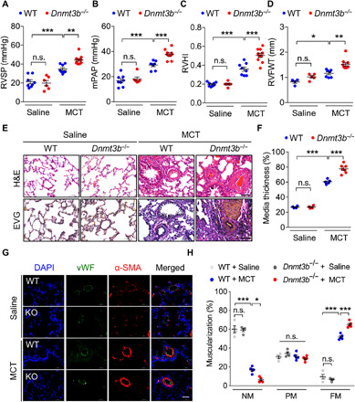
Fig. 2. Dnmt3b deficiency exacerbates pulmonary vascular remodeling in MCT-induced PH rat model.

(A to D) Compared to WT saline group, Dnmt3b−/− homozygous [Dnmt3b−/−, knockout (KO)] Sprague-Dawley rats at week 4 after MCT injection exhibited an elevation in (A) RVSP, (B) mPAP, (C) RVHI, and (D) right ventricular free wall thickness (RVFWT) (A to C, n = 8 for WT saline group, n = 6 for KO group, n = 8 for WT MCT group, and n = 10 for KO MCT group; D, n = 4 for WT saline group, n = 5 for KO group, n = 7 for WT MCT group, and n = 10 for KO MCT group). (E) Representative photomicrographs of hematoxylin and eosin (H&E) staining and elastin–van Gieson (EVG) staining of lung tissue from MCT or saline-treated rats at day 28. Original magnification, ×400. Scale bars, 50 μm. (F) Pulmonary vascular remodeling by percentage of vascular medial thickness to total vessel size (n = 4 to 7 per group) and (G) quantification of vessel muscularization by immunofluorescence staining with anti–α-SMA (red, smooth muscle cells), anti-vWF (green, endothelial cells), and 4′,6-diamidino-2-phenylindole (DAPI) (blue, nuclei) for the PH model, demonstrating that Dnmt3b deficiency promoted a further elevation of pulmonary vascular wall thickness after MCT injection. Scale bars, 50 μm. (H) Proportion of nonmuscularized (NM), partially muscularized (PM), or fully muscularized (FM) pulmonary arterioles (20 to 50 μm in diameter) from MCT-treated rats, confirming that Dnmt3b deficiency significantly increased arteriole muscularization (n = 4 to 5 per group). *P < 0.05, **P < 0.01, and ***P < 0.001 versus WT saline group or WT MCT group, one-way ANOVA with Bonferroni correction for multiple comparisons (A to D and F) and two-way ANOVA with Bonferroni’s post hoc analysis (H); mean ± SEM.
