
Fig. 6. Antiviral activity of GC-376 analogs.
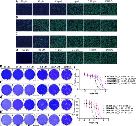
(A to D) Antiviral activity of GC-376 analogs against SARS-CoV-2 in the immunofluorescence assay. (A) GC-376; (B) UAWJ246; (C) UAWJ247; (D) UAWJ248. Vero E6 cells in a 96-well plate were infected with SARS-CoV-2 (USA-WA1/2020 isolate) at an MOI of 0.05 in the presence of the indicated concentrations of the tested compounds. At 48 hours post infection (hpi), the cells were fixed and stained with a rabbit monoclonal antibody against the SARS-CoV-2 NP and a secondary antibody conjugated with Alexa 488 (green). The nuclei were counterstained with Hoechst dye (blue). For each well, fluorescence images of approximately 10,000 cells were acquired and shown. The images are representatives of three repeats. (E to H) Antiviral activity of GC-376 analogs against SARS-CoV-2 in the plaque assay. (E) GC-376; (F) UAWJ246; (G) UAWJ247; (H) UAWJ248. Vero E6 cells in six-well plates were infected with approximately 40 plaque-forming units per well of SARS-CoV-2 (USA-WA1/2020 isolate). After 1 hour, the inoculum was removed, and the cells were overlaid with medium containing the indicated concentrations of the tested compounds and 1.2% Avicel RC-591. At 3 days post infection, the overlay was removed, and the cells were stained with 0.2% crystal violet. The images are representatives of two repeats. Data fitting of the antiviral activity of GC-376 analogs against SARS-CoV-2 in the immunofluorescence assay (I) and the plaque assay (J).