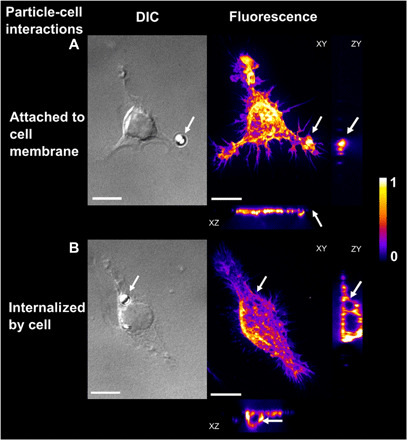
Fig. 1. Images of particle-cell interactions of microplastic particles exposed to fresh water for 2 weeks.

DIC: Differential interference contrast microscopy images of particle-cell interactions. Fluorescence: Spinning disc confocal images of the cells with fluorescently labeled filamentous actin (false color image, maximum intensity projection showing arbitrary units). XY, YZ, and XZ projections of three-dimensional confocal images allow the differentiation of microplastic particles (A) attached to cell membranes or (B) internalized microplastic particles. Arrows indicate microplastic particle position. Scale bars, 10 μm.
