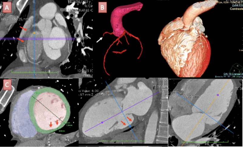
Figure 3. Coronary computed tomographic angiography findings of the patient.
A: Multiplane reconstruction of the right coronary artery (RCA) showing total occlusion at the proximal segment (red arrows). B: Tri-dimensional (3D) volume rendering of coronary arteries and cardiac volume showing total occlusion of the RCA (white arrows) and distribution of coronary arteries from an anteroposterior view of the heart. C: Multiple planes reconstruction of the left ventricle (LV). LV short-axis view (bottom left) estimated an end-diastolic LV diameter of 70 mm consistent with a severe dilatation and denoted an inferobasal wall thickness of 3.6 mm (red arrows). LV long-axis two and four-chamber views (bottom middle and right) consistent with inferior basal LV thinned wall (red arrows).

