Figure 3.

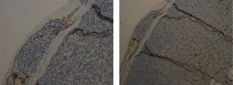
Section of the right inferior parathyroid specimen showing a venous vascular invasion focus highlighted by immunohistochemical staining for CD31 ((a) 20x; (b) 10x).

Section of the right inferior parathyroid specimen showing a venous vascular invasion focus highlighted by immunohistochemical staining for CD31 ((a) 20x; (b) 10x).