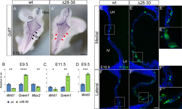
Fig. 8.
The mutant hChP shows changes in markers of roof-plate patterning. (A,A′) Dorsal view of whole-mount in situ hybridization reveals a conserved but discontinuous Gdf7 expression pattern in the mutant (A′, red arrows). (B) RT-qPCR quantitation of roof-plate patterning markers at E9.5 reveals deregulated expression of three early patterning molecules involved in Wnt and BMP signaling. (C) Deregulation of Wnt1 and Grem1 persists at E11.5. (D) Wnt3 is strongly upregulated at E9.5. Data are mean±s.e.m. (B,D, n=3/genotype; C, n=4/genotype). ****P<0.0001, ***P<0.0005, **P<0.005, *P<0.05 (Welch's unequal variances t-test). (E-F‴) Parasagittal cerebellar sections at E10.5 stained for p-ERK. Both medial (E-E‴) and lateral (F-F‴) sections show an increase of p-ERK expression in the mutant hChP primordium (box in E′,F′, magnified in E‴,F‴) compared with the wild type (box in E,F; magnified in E‴,F‴). Cb, cerebellum; Mes, mesencephalon; Rp, roof plate; Url, upper rhombic lip; Lrl, lower rhombic lip; IV, fourth ventricle. Scale bars: 500 μm in A,A′; 50 μm in E-F′; 120 μm E″-F‴.

