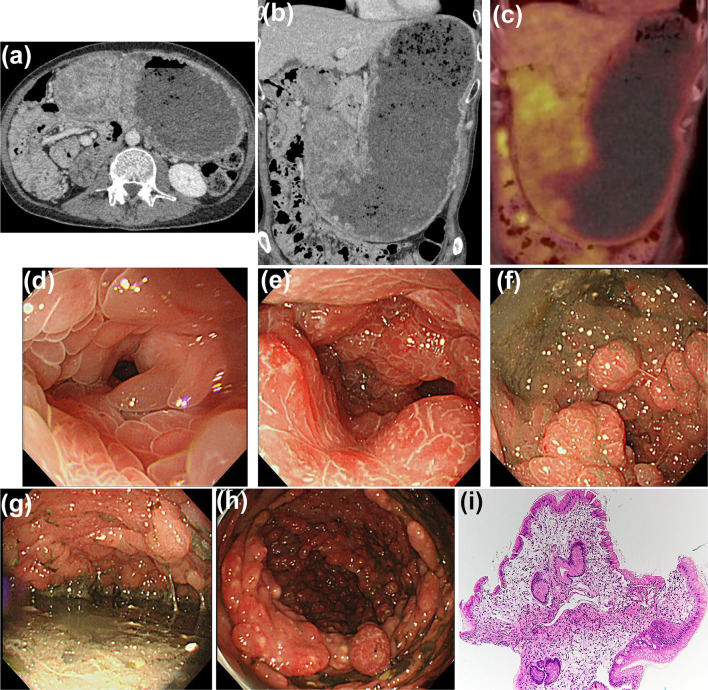
Figure 2.
Findings at CCS relapse. CT revealed massive gastric dilatation and marked wall thickening (a, b). No uptake of intense FDG was observed in the gastric wall thickening (c). EGD revealed gastric outlet obstruction due to the numerous edematous and reddish polyps (d-g). Furthermore, TCS revealed multiple reddish polyps along the entire colon (h). Histopathology of biopsied gastric polyp specimens demonstrated a substantially edematous stroma and inflammatory cell infiltration without malignancy (i, H&E, ×40). CCS: Cronkhite-Canada Syndrome, CT: computed tomography, FDG: fluorodeoxyglucose, EGD: esophago-gastroduodenoscopy, TCS: total colonoscopy, H&E: Hematoxylin and Eosin staining

