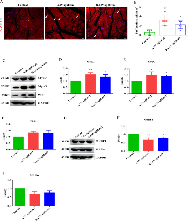
Fig. 5. Satellite cells are increased in number and myogenic proteins changed during the Mstn knockout in the left thigh of aged C57BL/10 male mice.
a Satellite cells expression. Immunofluorescent staining of muscle sections using a Pax7 antibody (in red). Representative images are shown. The nuclei were stained with DAPI (in blue). The white arrow indicates the satellite cell. b Quantification of the number of Pax7-positive cells/mm2 from a. Data are means ± SEM from five muscles derived from five different aged mice per group. *: 0.01 < P < 0.05, **P < 0.01. Western blot analysis was also performed in order to determine the kinds of cell factors protein levels. c MyoD, MyoG and Pax7 were detected during the Mstn knockout. Normalized data were used for generating the d MyoD, e MyoG and f Pax7 graphs depicted. Data represent means ± SEM (the number of samples was 3 per group, and each sample was repeated three times). *: 0.01 < P < 0.05, **P < 0.01 when AAV-sgMstn2, RAAV-sgMstn2-treated were compared with AAV-CN-treated. g Western blot was used to detect the effect of MSTN on the expression of MuRF1 and MAFbx proteins. Normalized data were used for generating the h MuRF1, i MAFbx graphs depicted. Data represent means ± SEM (the number of samples was 3 per group, and each sample was repeated three times). *: 0.01 < P < 0.05, **P < 0.01 when AAV-sgMstn2, RAAV-sgMstn2-treated were compared with AAV-CN-treated.

