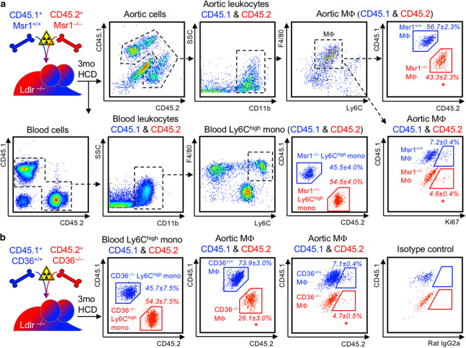
Fig. 5.
Cholesterol-rich modified LDL-uptake-mediating scavenger receptors Msr1 and CD36 directly propagate MF proliferation in plaques. a Ldlr–/– mice were irradiated and reconstituted with a 1:1 mixture of CD45.1 Msr1+/+ and CD45.2 Msr1–/– bone marrow cells for 6 weeks before starting a high-cholesterol diet (HCD) for 3 months. Msr1+/+ and Msr1–/– Ly6Chigh monocytes (mono) in blood and macrophages (Mϕ) in enzymatically digested atherosclerotic aortas were distinguished based on exclusive CD45.1 and CD45.2 expression, as depicted in the representative dot plots and gating strategies. The chimerism of Msr1+/+ and Msr1–/– within blood Ly6Chigh monocytes and aortic Mϕ was quantified based on CD45.1 and CD45.2 staining, and the fraction of proliferating cells was assessed based on intracellular Ki67 staining in Msr1+/+ and Msr1–/– MF. Results are presented as mean percent ± SEM cell chimerism and Ki67+ fraction of the respective population, n = 4 per group, *p < 0.05 denotes statistically significant differences between Msr1+/+ and Msr1–/– Ly6Chigh monocyte or Mϕ population, Mann–Whitney test. b Mixed CD45.1 CD36+/+ and CD45.2 CD36–/–irradiation bone marrow chimeras were generated in Ldlr–/– mice and analyzed in analogy to Msr1+/+/Msr1–/– chimeras, as described above. Results are presented as mean percent ± SEM cell chimerism and Ki67+ fraction of the respective population, n = 4 per group, *p < 0.05 denotes statistically significant differences between CD36+/+ and CD36–/– Ly6Chigh monocyte or Mϕ population, Mann–Whitney test

