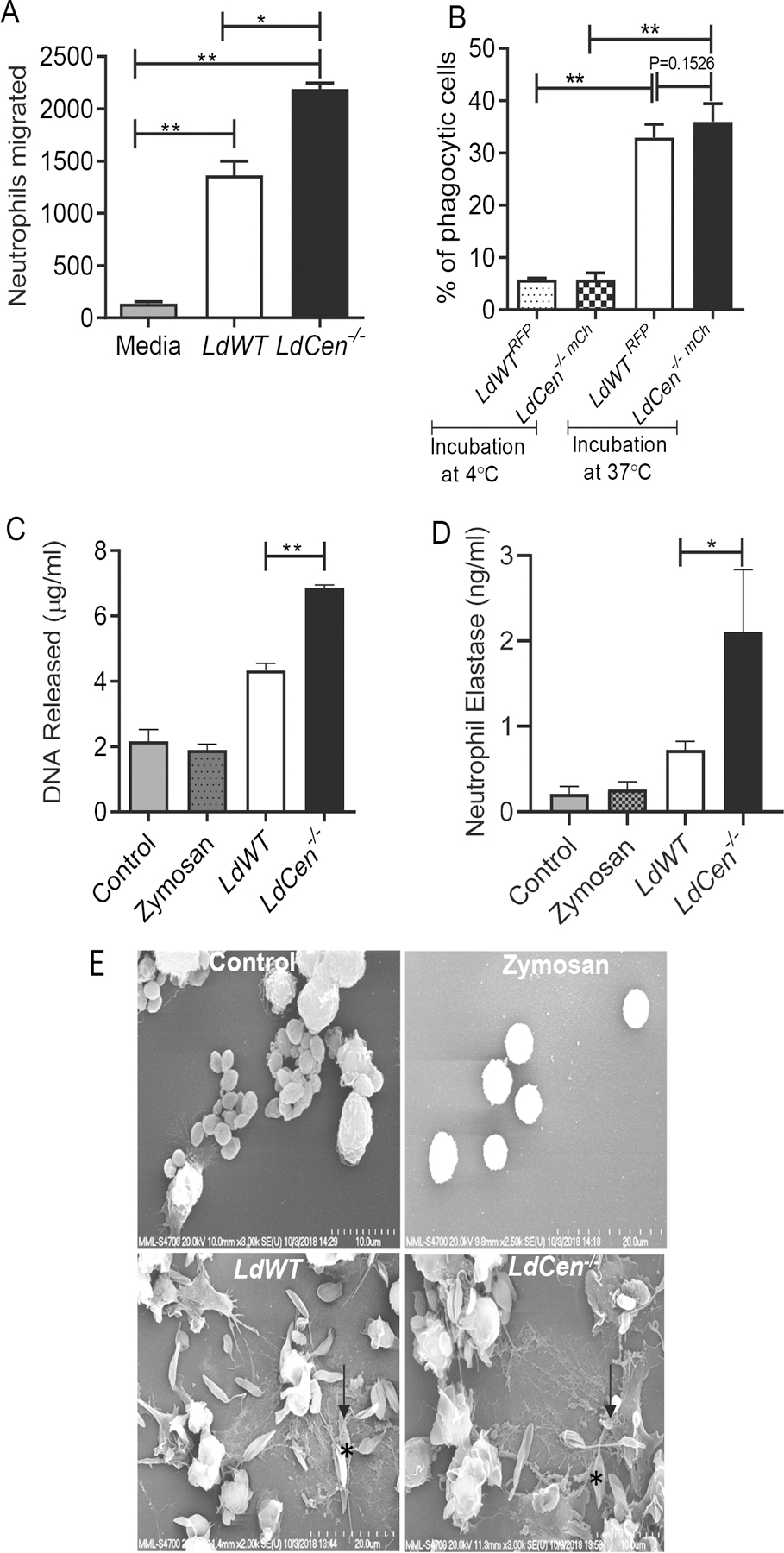
Figure 1: LdCen−/− infection induced strong effector function in neutrophils compared to LdWT infection in vitro.

(A) Absolute number of neutrophils that migrated after stimulation with supernatants of LdWT or LdCen−/− infection (6h) of macrophages was measured by chemotaxis assay. The data represent the mean values ± standard deviations (SD) of results from 3 independent experiments that all yielded similar results. * P < 0.05; ** P < 0.005. (B) Peritoneal neutrophils were cocultured with LdWTRFP or LdCen−/−mcherry for 4h (1:5 cell: parasite ratio) either at 4℃ or at 37℃. Bar diagrams represent percentages of RFP+/mCherry+ neutrophils as determined by flow cytometry. Data are pooled from 3 independent repeats and are shown as means ± standard deviations (** P < 0.005) between the groups. (C) Neutrophils were incubated either with LdWT or LdCen−/− promastigotes or with zymosan. DNA released was quantified after 6h of incubation. The data presented are means ± SD of 3 independent experiments. ** P < 0.005 between the groups. (D) Peritoneal neutrophils were lysed and assayed for NE activity as described in Material Methods. The NE activity was expressed as the absorbance observed at 450 nm. Data are pooled from 3 independent repeats and are shown as means ± standard deviations (*P < 0.05) between the groups. (E) Scanning electron microscopy for visualization of NET. Naïve neutrophils were either remain uninfected or incubated with zymosan or promastigotes (as indicated by*) for 6h. NET fibers (as indicated by black arrow) were found in infected (LdWT or LdCen−/− ) neutrophils. The micrographs are representative of 3 independent experiments in which at least 100 cells per sample were analyzed.
